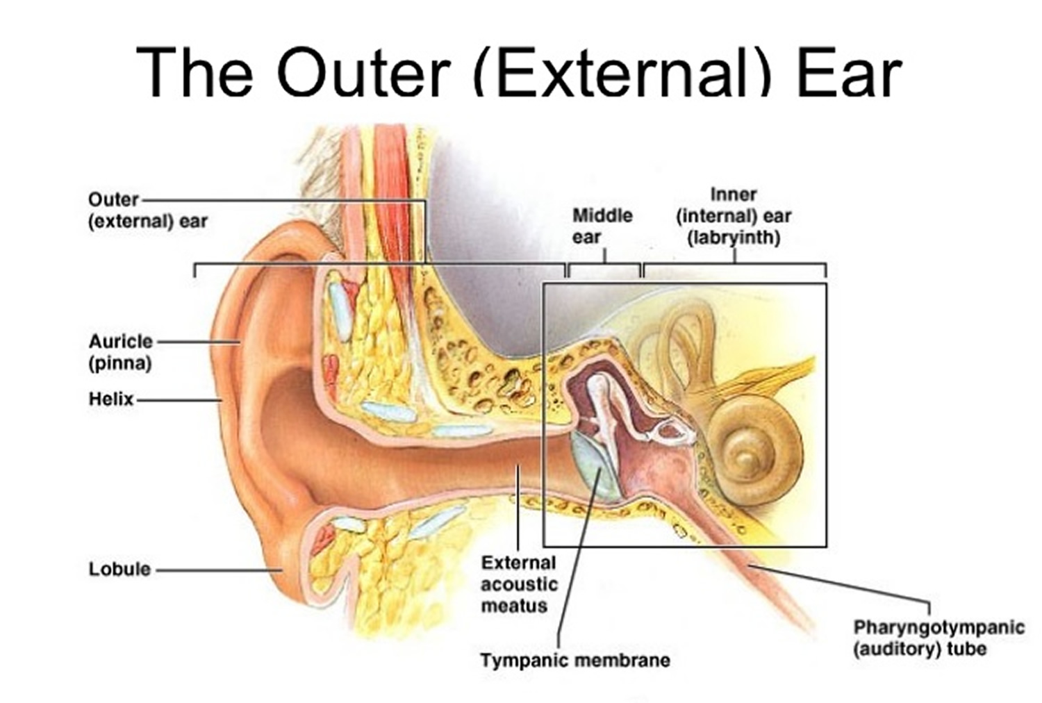
The inner ear is probably the most complicated part of the human body. It enables hearing by converting sound into electrical signals which then travel along the auditory nerve to the brain. The inner ear also plays a role in balance. The parts involved in balance (membrane maze) can detect the acceleration of the head in any direction, either in a straight line (linear acceleration) or a rotation or turn (angular acceleration). The electrical signals which are generated in response to movements of the head pass through the nerve of balance (vestibular nerve), which in its course combines with the cochlear nerve to form a single bundle (the auditory nerve), which then enters the brain.

The part of the inner ear that actually “listens” is the cochlea. This is a hollow, helical tube in very dense bone, called bone labyrinth (part of part of the lithoeidi fate of the temporal tissue). The tube is filled with fluid similar to that which circulates throughout the body (lymph) and the fluid surrounding the brain (cerebrospinal fluid). This internal fluid is called outer lymph. Inside the outer lymph is another spiral, triangular-shaped tube called a cochlear duct, which contains the very important “hair cells” (they convert sound into electrical current). These hair cells are arranged in two groups following the cochlear duct spirals upward from the bottom. There is a single row of inner hair cells near the body of the cochlia and 3-4 rows of outer hair cells. In healthy young humans there are about 3,500 inner hair cells and about 12,000 outer hair cells.
Each hair cell has a group of small powerful cilia (stereokrossoi), which emerge from the thicker upper surface of the cell in a special fluid that fills the cochlear duct. The fluid is called inner lymph and what is remarkable is that it is characterized by a strong positive electric charge (about 80 millivolt) and the fact that it is rich in potassium, a mineral element.
The hair cells are arranged together with their supporting cells within the organ of Corti. This is a thin ridge which lies on a thin, highly flexible membrane, which in turn is called the main membrane. The main membrane forms the territory of the triangular cochlear duct. The downward roof is formed by another thin membrane (vestibular membrane) and the lateral wall by a thickened area rich in blood vessels (vascular tape). This structure is responsible for maintaining the composition of the unusual but very significant inner lymph.
In contact with the base of the hair cells are nerve fibers that carry impulses (signals) to the brain (afferent fibers). At least 90% of these nerve fibers start from the inside cells, despite their lower numbers. Every inner hairy cell has about 10 nerve endings that are attached to it, and this means that there are about 30,000 nerve fibers in the cochlear nerve.
Outer hair cells have nerve fibers associated with them, but most of them are fibers coming from the brain (efferent fibers).