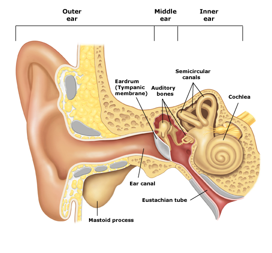
The tympanic membrane is a circular disk of thin skin with a diameter 8-9mm. Unlike its name, it is not flat like the surface of a music drum, but has a rather conical shape with its apex inward. The middle ear itself is located in the inner membrane of the tympanic membrane and is a ventilated area which contains three small bones (ossicles) that connect the tympanic membrane to the inner ear.

The ossicles called malleus, incus and stirrup because of their similarity to these objects. The malleus has a handle and a head and the handle is positioned between the layers of the tympanic membrane. The head of the hammer abuts an area of the middle ear and is hinged (the joint is like any other joint in the body) on the rather chunky body of the anvil. From the anvil starts a long appendage directed toward the bottom wall of the middle ear and eventually connects with the head of the stirrup. The two arms of the stirrup are connected at the base of the stirrup which is inside a small hole (3 mm x 2 mm) called the oval window. This is the opening to the fluid-filled space of the inner ear. Just below the oval window there is another small hole towards the inner ear called the round window. A thin membrane covers this hole and when the base of the stirrup moves “inside-out” the membrane of the round window moves “outside-in” because the fluid in the inner ear transmits pressure changes.
Through the middle ear moves the facial nerve (seventh cranial nerve). The nerve leaves the brain and passes through the skull to get to the muscles of the facial expression, i.e. the frown muscles, the smile muscles etc. The nerve is located in a thin bony canal and runs horizontally from the front to the rear of the middle ear, just above the oval window and stirrup, before turning downward, leaving the base of the skull. Subsequently, the nerve is directed forwards to reach the face. Therefore, the facial nerve is sensitive to lesions of the middle ear and to the risks of injury from the surgery of the middle ear. The paralysis of the facial nerve leads to paralysis of one side of the face, resulting in the fall of the face and the impossibility of motion. The smile leads to grimace, swallowing liquids in their loss from the mouth, while the eye cannot close.
In the middle ear there are two small muscles. One of them is in the front (called tends the drum muscle) adhered to the top of the handle of the hammer and stretches the drum when activated by ingestion. The function of this muscle is unclear, but probably aims at making the feeding and swallowing less noisy.
The muscle located in the rear of the middle ear (called the stapedius muscle) begins near the facial nerve, from where it is innervated and adheres to the head of the stirrup. Reacts to loud sounds with contraction and tightening of the chain of the ossicles and probably reduces the transmission of prolonged and potentially damaging sounds to the inner ear.
The middle ear is therefore a ventilating area coated with active tissue that is able to produce debris from dead epithelial cells as well as mucus from the glands. This creates two problems: first, the need for cleaning debris and mucus and a second less obvious but very important problem. The oxygen in the air of the middle ear is absorbed by blood vessels at the mucosa in the same way that oxygen is absorbed by the lungs. A quantity of carbon dioxide is released from these vessels into the air of the middle ear, but the overall effect is the pressure drop in the middle ear, since more oxygen is removed in relation to the carbon dioxide that is produced. With the atmospheric pressure prevailing outside the tympanic membrane, something of the whole structure will necessarily succumb and the only part that can be moved is the tympanic membrane. That could be moved inwards and then cease to function normally. Eventually, the entire middle ear would collapse and would cause a significant decrease in hearing.
When operating normally, the auditory (Eustachian) tube prevents the development of these problems. Its course is towards the front and inside, starting from the anterior wall of the middle ear to reach the rear of the nasal cavity above the soft palate (the Nasopharynx). The opening near the tip is soft and pliable and opens during swallowing and yawning. Although we do not know exactly how this mechanism works, when the Eustachian tube opens, enough air enters the middle ear to replenish the oxygen that has been absorbed and to keep the pressure of the middle ear at levels close to the atmospheric pressure. It has been estimated that 1-2 ml of air per ear every day, that is less than half a teaspoon of air, is sufficient to maintain adequate ventilation of the middle ear, but without it, the middle ear cannot function normally.
The Eustachian tube is also the channel through which, by means of cilia, the mucus produced in the middle ear to the back of the nose is being removed, where it can be swallowed. This thin layer of mucus, which also transfers all residues produced in the middle ear, moves in the territory of the Eustachian tube, with the air directed through the nose to the middle ear moving over it. Thus, both ventilation and self-cleaning are achieved when the system is working properly. Unfortunately, in humans this system is relatively sensitive and often not working sufficiently, probably due to the shape of the skull needed host the large brain.
There is another extension of the ventilated spaces of the middle ear space towards the rear, in the mastoid bone. You can feel this as a circular shape if you put your hand on the back of your ear. The mastoid bone should be concave, with its ventilating spaces separated by small and incomplete bone septa, reminiscent of cell morphology. An average mastoid antrum contains an air volume of about 15-20 ml (3-4 teaspoons) and serves to balance the pressure changes in the middle ear and to reduce the adverse effects on the tympanic membrane. People with small mastoid cells appear to have higher risk of developing diseases of the middle ear and mastoid antrum. At present it is unknown whether the disease of the middle ear or mastoid antrum is responsible for growth inhibition of the antrum or if the small size of the mastoid antrum decreases the balancing of the pressure and causes the development of the disease. The probable answer is that it can be a combination of both.